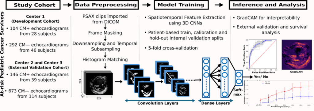
We’re pleased to share a new paper led by Surbhi Sharma, in collaboration with Lindsay Edwards (Duke) and colleagues across Seattle Children’s, Fred Hutch, and other children’s hospitals: “Early prediction of cardiomyopathy in pediatric cancer survivors using echocardiographic video-based deep learning” (also available on PubMed Central).
Cardiomyopathy (CM) is a leading cause of late death in childhood cancer survivors, but it is often caught only after irreversible cardiac remodeling. We asked whether a 3D convolutional neural network, applied to routine parasternal short-axis echo clips, could pick up subtle signs of CM up to five years before diagnosis.
Key findings:
- On an internal hold-out set from one center, the model reached an AUROC of 0.87 and an AUPRC of 0.74.
- Performance on two external centers was much weaker for single-study classification (AUROC 0.55 and 0.60).
- Even so, model-defined high-risk survivors in the external cohort were more likely to develop CM over time than low-risk survivors (hazard ratio 2.78, 95% CI 1.52 to 5.10).
This is a proof-of-concept study, and the drop in external performance shows how much work remains on generalizability across institutions. We see it as a first step toward risk-guided surveillance that complements, rather than replaces, current care.
This work was supported by the NIH, the Rally Foundation, the Catherine Holmes Wilkins Charitable Foundation, the Unravel Pediatric Cancer Foundation, the Seattle Foundation, and the Seattle Children’s Research Institute.